Needle Biopsy (Image guided)
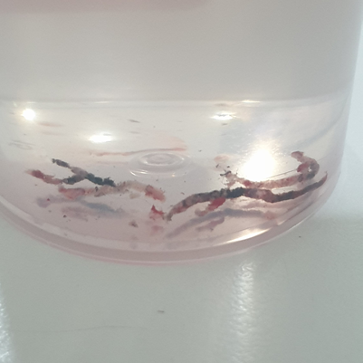
A needle biopsy is a common, minimally invasive procedure used to obtain a small sample of tissue (from a lump or abnormal area) for examination under a microscope. This is often necessary to determine the exact cause of an abnormality seen on an imaging test.
What are the Benefits? (Why have the procedure?)
The main benefit of a needle biopsy is to provide a definitive diagnosis with high accuracy without the need for open surgery. This is critical for planning the right treatment.
- Accurate Diagnosis: Determines if an abnormality is benign (non-cancerous), malignant (cancerous), or caused by an infection or other condition.
- Minimally Invasive: Uses only a small needle, avoiding the larger incision and general anesthesia required for surgical biopsy.
- Faster Recovery: Patients typically return to normal activities much sooner than after surgery (often the next day)
- Treatment Planning: If cancer is found, the tissue provides vital information (like tumor type and characteristics) that helps your medical team choose the most effective treatment plan.
Associated Risks and Complications
Needle biopsies are generally safe procedures, but like any medical procedure, there are potential risks and complications.
| Bleeding/Bruising | Minor bleeding at the biopsy site is common. Significant bleeding that requires intervention is rare. | Low (Clinically significant bleeding is often in many studies). |
| Infection | The risk of infection is very low due to sterile techniques. Antibiotics may be given if an infection occurs. | Very Low (Generally or very rare). |
| Pain/Soreness | Discomfort, pain, or soreness at the site is common and usually managed with over-the-counter pain relievers. | Common, but usually mild and temporary. |
| Pneumothorax (Collapsed Lung) | Primarily a risk for lung biopsies. Air leaks into the space between the lung and chest wall. This is the most common complication of a lung biopsy, but often resolves on its own. | Moderate ( for any pneumothorax after lung biopsy); Low (Only about require a chest tube). |
| Inconclusive Result | In a small number of cases, not enough tissue is collected, or the sample is not adequate for a definitive diagnosis. This may require a repeat biopsy or a surgical biopsy. | Low (Varies by lesion and technique, typically of image-guided biopsies). |
| Injury to Nearby Structures | Very rare risk of damage to a nerve, blood vessel, or an adjacent organ/structure. | Very Rare (Highly minimized by imaging guidance). |
Note: Specific risks can vary based on the organ being biopsied (e.g., the risk of pneumothorax is specific to lung biopsy). Your doctor will discuss risks specific to your procedure.
Imaging Guidance: How We Ensure Accuracy
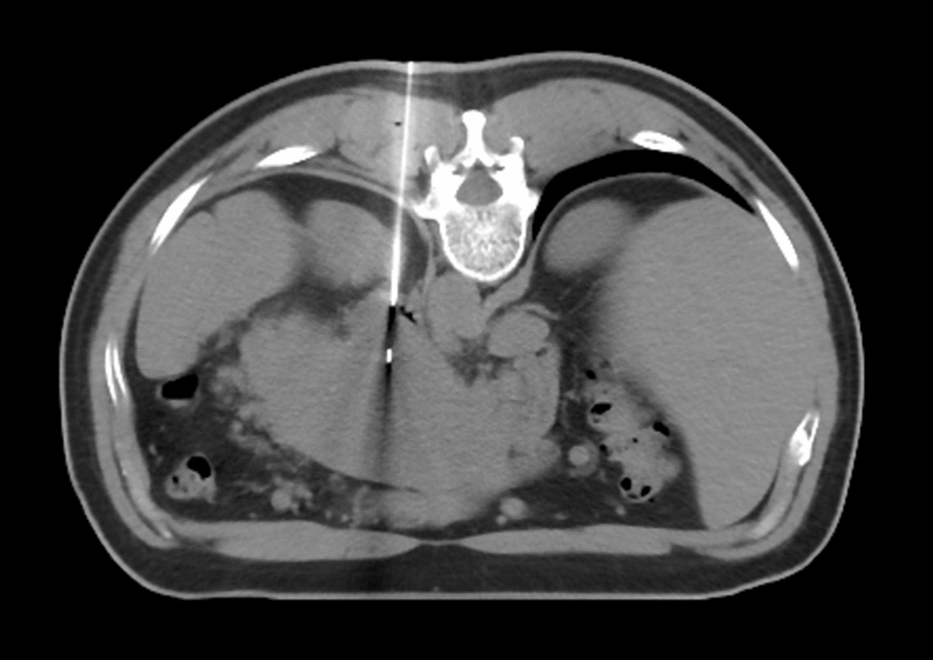
A needle biopsy is an image-guided procedure. This means the doctor uses real-time imaging technology to see inside your body, pinpoint the exact location of the abnormal tissue, and guide the needle safely to the target.
| Imaging Guidance | When It’s Typically Used |
| Ultrasound (US) | Often used for easily accessible lesions, such as those in the breast, thyroid, liver, or enlarged lymph nodes. It provides real-time visibility for needle movement. |
| Computed Tomography (CT) Scan | Used for deeper lesions, such as those in the lung, abdomen, bone, or other areas not clearly seen on ultrasound. It provides a detailed, cross-sectional view. |
| Magnetic Resonance Imaging (MRI) | Primarily used for abnormalities best seen on MRI, such as certain breast lesions or prostate tissue. |
| Stereotactic Mammography | Used for breast lesions (like microcalcifications) that are only visible on a mammogram. It uses X-ray images from multiple angles to map the exact target location in three dimensions. |
Sources: